An Aston University researcher has developed a new technique using light that could revolutionize non-invasive medical diagnostics and optical communication. The research showcases how a type of light called the orbital angular momentum (OAM) can be harnessed to improve imaging and data transmission through skin and other biological tissues.
A team led by Professor Igor Meglinski found that OAM light has unmatched sensitivity and accuracy that could result in making procedures such as surgery or biopsies unnecessary. In addition, it could enable doctors to track the progression of diseases and plan appropriate treatment options.
OAM is defined as a type of structured light beams, which are light fields which have a tailored spatial structure. Often referred to as vortex beams, they have previously been applied to a number of developments in different applications including astronomy, microscopy, imaging, metrology, sensing, and optical communications.
Professor Meglinski, in collaboration with researchers from the University of Oulu, Finland, conducted the research, which is detailed in the paper “Phase preservation of orbital angular momentum of light in multiple scattering environment” published in Light: Science & Applications. The paper has since been named as one of the year’s most exciting pieces of research by the international optics and photonics membership organization, Optica.
The study reveals that OAM retains its phase characteristics even when passing through highly scattering media, unlike regular light signals. This means it can detect extremely small changes with an accuracy of up to 0.000001 on the refractive index, far surpassing the capabilities of many current diagnostic technologies.
Professor Meglinski, who is based at Aston Institute of Photonic Technologies, said, “By showing that OAM light can travel through turbid or cloudy and scattering media, the study opens up new possibilities for advanced biomedical applications.
“For example, this technology could lead to more accurate and non-invasive ways to monitor blood glucose levels, providing an easier and less painful method for people with diabetes.”
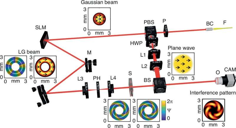
The research team conducted a series of controlled experiments, transmitting OAM beams through media with varying levels of turbidity and refractive indices. They used advanced detection techniques, including interferometry and digital holography, to capture and analyze the light’s behavior. They found that the consistency between experimental results and theoretical models highlighted the ability of the OAM-based approach.
The researchers believe that their study’s findings pave the way for a range of transformative applications. By adjusting the initial phase of OAM light, they believe that revolutionary advancements in fields such as secure optical communication systems and advanced biomedical imaging will be possible in the future.
Professor Meglinski added, “The potential for precise, non-invasive transcutaneous glucose monitoring represents a significant leap forward in medical diagnostics.
“My team’s methodological framework and experimental validations provide a comprehensive understanding of how OAM light interacts with complex scattering environments, reinforcing its potential as a versatile technology for future optical sensing and imaging challenges.”
More information:
Igor Meglinski et al, Phase preservation of orbital angular momentum of light in multiple scattering environment, Light: Science & Applications (2024). DOI: 10.1038/s41377-024-01562-7
Provided by
Aston University
Citation:
Optical technique that uses orbital angular momentum could transform medical diagnostics (2024, October 25)
retrieved 25 October 2024
from https://phys.org/news/2024-10-optical-technique-orbital-angular-momentum.html
This document is subject to copyright. Apart from any fair dealing for the purpose of private study or research, no
part may be reproduced without the written permission. The content is provided for information purposes only.

